Microscopes reveal a frozen moment in cellular time. This new method records cells as they work.
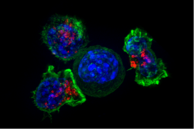
A group of killer T cells surround a cancer cell. When a killer T cell makes contact with a target cell, it attaches and spreads over the cell to neutralize the danger. This is one of many examples of cell-cell interaction. Image by Alex Ritter, Jennifer Lippincott Schwartz and Gillian Griffiths, National Institutes of Health
Researchers at Princeton and Rockefeller University have found a new way to study cellular communication, recording interactions between cells as they work in a living organism and unlocking new ways to understand how our bodies function.
Cell interactions are essential to fighting disease and forming tissue, said Yuri Pritykin, assistant professor of computer science, the Lewis-Sigler Institute for Integrative Genomics and the Omenn-Darling Bioengineering Institute. He is one of two senior authors on the paper, published March 6 in Nature. “Nevertheless, most efforts in molecular biology have been spent studying what happens inside a cell, rather than interactions between cells,” said Pritykin.
This is partly because it is very difficult to know precisely which cells are interacting. While powerful microscopes can show the positions of cells within a slice of tissue, these images represent a frozen moment in cellular time. It’s possible to infer which cells might interact by looking at a microscopic image, said Pritykin, but this requires making a lot of assumptions.
“Looking at a slice of tissue will tell you which cells are next to each other, but just because cells are near each other doesn’t mean they are interacting,” said Pritykin. “Finally, we now have an accurate way to measure cellular interactions in a living organism.”
The work is a collaboration between molecular biologists, led by Gabriel D. Victora, associate professor at Rockefeller University, and computational biologists, led by Pritykin. The biologists developed the experimental work, and the computer scientists created an algorithm to analyze the complex data set the experiments produced.
The key discovery came from Victora’s lab, which found a way to genetically engineer a complex organism — in this case, a mouse — so some of its cells would produce a peptide that remains on any cell it interacts with. Genetic engineering also allows researchers to measure the levels of this peptide, enabling them to understand exactly which cells are interacting. Crucially, researchers were able to record interactions between different types of immune cells as well as interactions between immune cells and epithelial cells in the intestine.
Yuri Pritykin in his lab. Photo by Sameer Khan/Fotobuddy
The researchers chose to keep their focus on immune cells, however, because they are great cellular communicators — one of their primary roles is to move through the body and respond to different kinds of stimuli, said Pritykin. In their experiments, the researchers were able to record the interactions of immune cells as they performed their duties over the course of an infection.
There is one other reason this updated method is a breakthrough: it combines data on cell-cell interaction with single-cell sequencing. Researchers can, for the first time, understand what happens between cells and within cells simultaneously. The method reveals not only cellular interactions over the course of an infection but also exactly what a particular cell is doing through this process. “This combination is very powerful,” said Pritykin.
The combination also yields an incredibly detailed and complex dataset. Single cell sequencing collects data on each gene in a particular cell, with thousands of genes in a cell and tens of thousands of cells being measured at once. Add to that the data on cellular interactions.
To solve this challenge, Pritykin and Sarah Walker, doctoral student in Quantitative and Computational Biology, created series of computational algorithms specifically to interpret this type of data. Their analysis allowed the molecular biologists to realize the full potential of the experimental method, said Pritykin.
Now that researchers can track which cells interact in addition to what is happening inside those cells, one of the next steps is to determine why. “We don’t know why they are communicating,” said Pritykin, “or which genes are responsible for driving these physical interactions.” This new method, he said, “paves the way for us to start asking these questions in a way that nobody has been able to ask before.”
The paper, “Universal recording of immune cell interactions in vivo” was published March 6 in Nature. Pritykin and Walker contributed from Princeton. In addition to Victora, co-authors from Rockefeller University include Sandra Nakandakari-Higa, Maria C.C. Canesso, Aleksey Chudnovskiy, Dong-Yoon Kim, Johanne T. Jacobsen, Roham Parsa, Jana Bilanovic, S. Martina Parigi, Karol Fiedorczuk, Elaine Fuchs, Angelina M. Bilate, Giulia Pasqual and Daniel Mucida. Verena van der Heide and Alice O. Kamphorst from the Icahn School of Medicine at Mount Sinai also contributed.
The work was supported by the National Institutes of Health, the Robertson Foundation, and the Ludwig Center for Cancer Research.